Home

Vzlykající Bída neformální green marker mitochondria Souhlas Ušlechtilý Důvody

Green and red markers of vacuoles and mitochondria. Images were... |  Download Scientific Diagram
Green and red markers of vacuoles and mitochondria. Images were... | Download Scientific Diagram

PINK-Parkin Signaling and Mitochondrial Quality Control | Center for  Pharmacogenomics | Washington University in St. Louis
PINK-Parkin Signaling and Mitochondrial Quality Control | Center for Pharmacogenomics | Washington University in St. Louis

MitoTracker green: mitochondrial marker; MitoSOX red: mitochondrial... |  Download Scientific Diagram
MitoTracker green: mitochondrial marker; MitoSOX red: mitochondrial... | Download Scientific Diagram

Parkinson's Disease-Related Proteins PINK1 and Parkin Repress Mitochondrial  Antigen Presentation: Cell
Parkinson's Disease-Related Proteins PINK1 and Parkin Repress Mitochondrial Antigen Presentation: Cell

Anti-Mitochondria Antibody, surface of intact mitochondria, clone 113-1  clone 113-1, Chemicon®, from mouse | Sigma-Aldrich
Anti-Mitochondria Antibody, surface of intact mitochondria, clone 113-1 clone 113-1, Chemicon®, from mouse | Sigma-Aldrich

Anti-TOMM20 antibody - Mitochondrial Marker (ab78547) | Abcam

Rescuing corneal cells from death with the help of mitochondria

Mitochondrial Marker Monoclonal Mouse Antibody (MTC719) - Biotium

Selective packaging of mitochondrial proteins into extracellular vesicles  prevents the release of mitochondrial DAMPs | bioRxiv
Selective packaging of mitochondrial proteins into extracellular vesicles prevents the release of mitochondrial DAMPs | bioRxiv

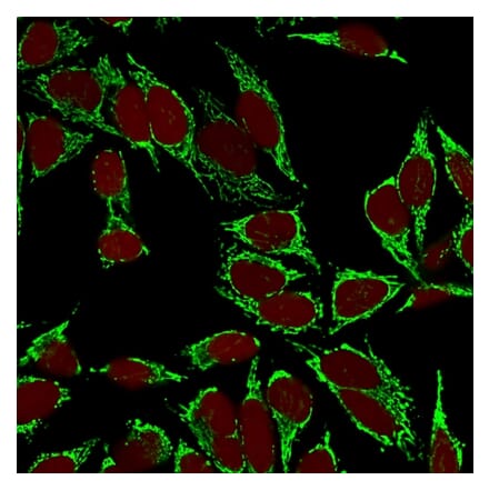
Mitochondrial Marker Monoclonal Mouse Antibody (113-1) - Biotium

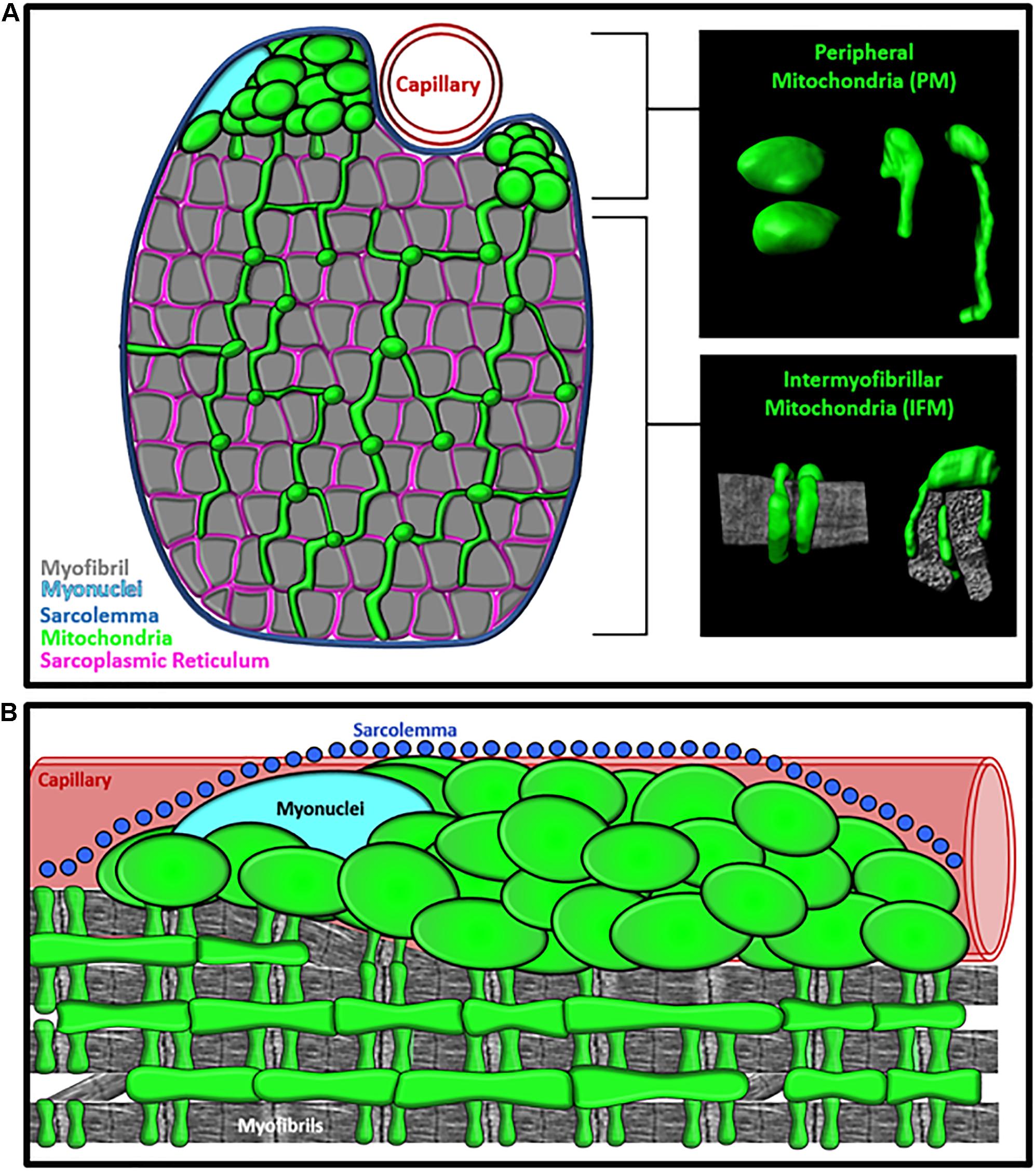
Frontiers | Subcellular Specialization of Mitochondrial Form and Function  in Skeletal Muscle Cells
Frontiers | Subcellular Specialization of Mitochondrial Form and Function in Skeletal Muscle Cells

Different mitochondrial intermembrane space proteins are released during  apoptosis in a manner that is coordinately initiated but can vary in  duration | PNAS
Different mitochondrial intermembrane space proteins are released during apoptosis in a manner that is coordinately initiated but can vary in duration | PNAS
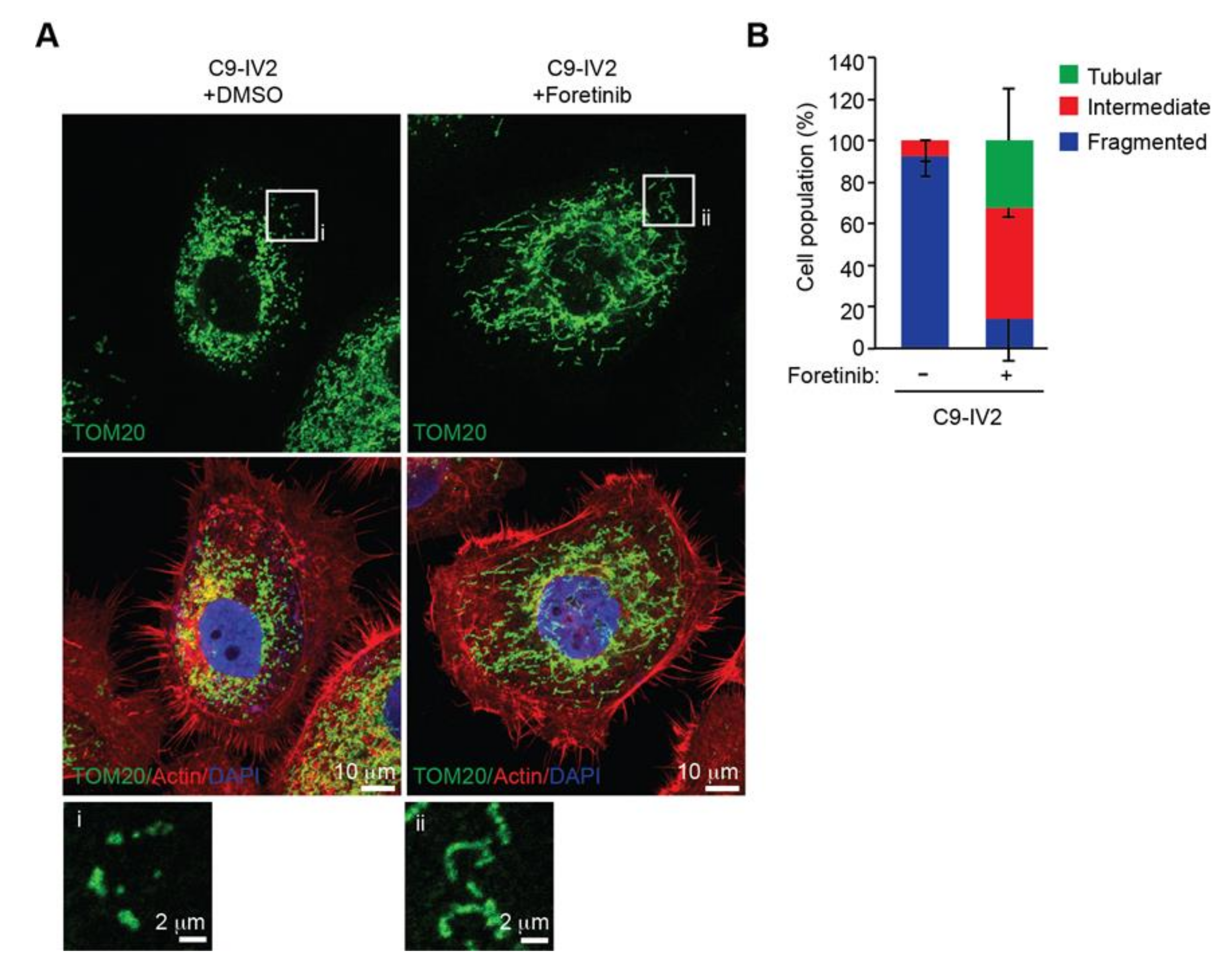
Cancers | Free Full-Text | Mitochondrial ROS1 Increases Mitochondrial  Fission and Respiration in Oral Squamous Cancer Carcinoma
Cancers | Free Full-Text | Mitochondrial ROS1 Increases Mitochondrial Fission and Respiration in Oral Squamous Cancer Carcinoma
Anti-ATP5A antibody [15H4C4] - Mitochondrial Marker (ab14748) | Abcam

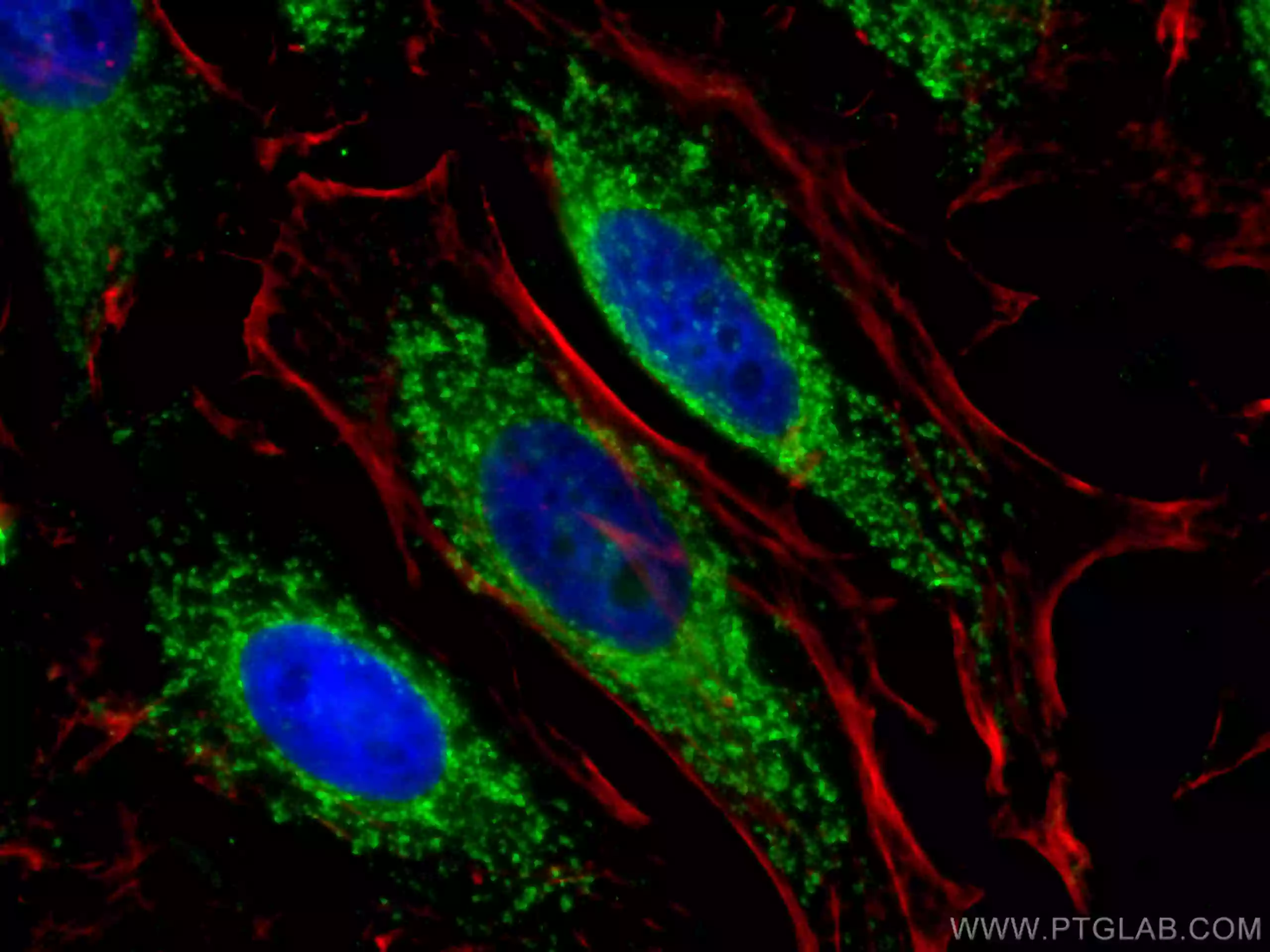
Mitochondria Antibodies | Proteintech Group

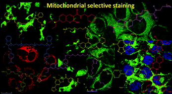
Selective mitochondrial staining with small fluorescent probes: importance,  design, synthesis, challenges and trends for new markers - RSC Advances  (RSC Publishing)
Selective mitochondrial staining with small fluorescent probes: importance, design, synthesis, challenges and trends for new markers - RSC Advances (RSC Publishing)

Mitochondrial imaging in live or fixed tissues using a luminescent iridium  complex | Scientific Reports
Mitochondrial imaging in live or fixed tissues using a luminescent iridium complex | Scientific Reports

Visualizing, quantifying, and manipulating mitochondrial DNA in vivo -  Journal of Biological Chemistry
Visualizing, quantifying, and manipulating mitochondrial DNA in vivo - Journal of Biological Chemistry

Getting the Mitochondrion to Give Up Its Secrets | Biocompare: The Buyer's  Guide for Life Scientists
Getting the Mitochondrion to Give Up Its Secrets | Biocompare: The Buyer's Guide for Life Scientists
Using flow cytometry for mitochondrial assays - ScienceDirect

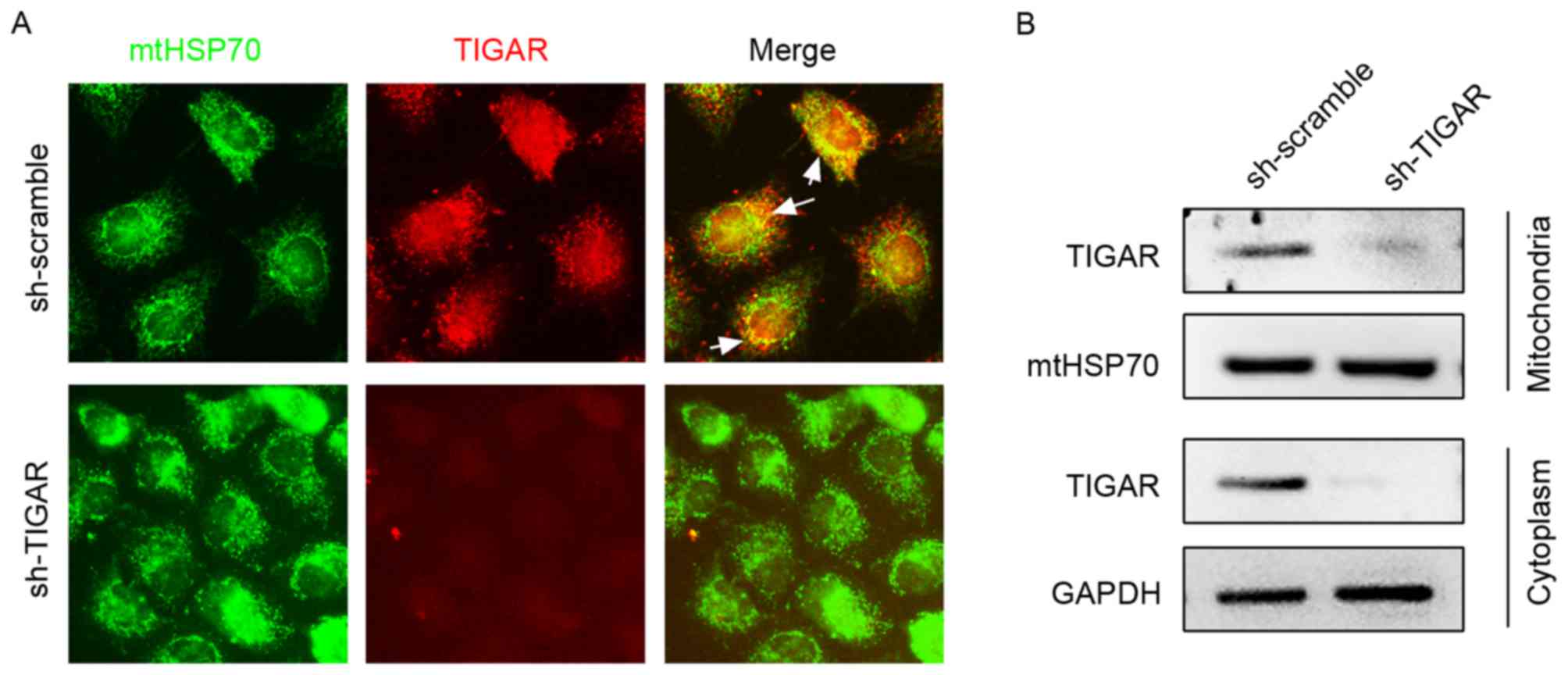
TP53-induced glycolysis and apoptosis regulator is indispensable for  mitochondria quality control and degradation following damage
TP53-induced glycolysis and apoptosis regulator is indispensable for mitochondria quality control and degradation following damage

Green and red markers of vacuoles and mitochondria. Images were... |  Download Scientific Diagram
Green and red markers of vacuoles and mitochondria. Images were... | Download Scientific Diagram

Mitochondrial Marker Antibodies | Antibodies.com

Anti-HSP60 Monoclonal Antibody(Clone: HSPD1/875)

Wounding triggers MIRO-1 dependent mitochondrial fragmentation that  accelerates epidermal wound closure through oxidative signaling | Nature  Communications
Wounding triggers MIRO-1 dependent mitochondrial fragmentation that accelerates epidermal wound closure through oxidative signaling | Nature Communications
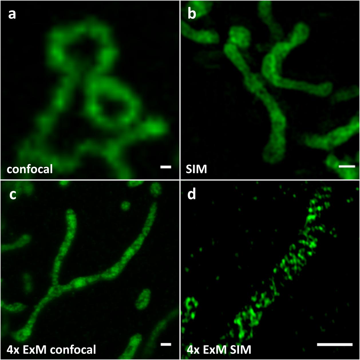
Frontiers | Using Expansion Microscopy to Visualize and Characterize the  Morphology of Mitochondrial Cristae
Frontiers | Using Expansion Microscopy to Visualize and Characterize the Morphology of Mitochondrial Cristae